ECG is basically the graphical representation of the electrical activity of cardiac muscles during contraction and release stages. It helps in determination of the cardiac arrhythmias in a well manner. Due to this early detection, arrhythmias can be done properly. In other words, we can say that the bio-potentials generated by the cardiac muscles results in an electrical signal called Electro-cardiogram (ECG). Feature extraction of ECG plays a vital role in manual as well as automatic analysis of ECG for the use in specially designed instruments like ECG monitors, Holter tape recorders and scanners, ambulatory ECG recorders and analyzers. In this paper, the study of pattern recognition of ECG is done. The ECG signal generated waveform gives almost all information about activity of the heart. The feature extraction of ECG is by Wavelet transform. This paper also includes artificial neural network as a classifier for identifying the abnormalities of heart disease.
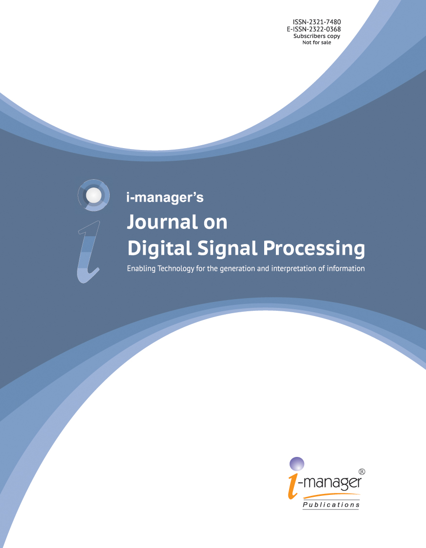
Electrocardiography gives information of the electrical activity of the cardiac muscles. Bio-signals which are usually non-stationary signals may occur randomly in the time-scale. Hence, for the effective diagnosis, the ECG signal pattern and heart rate variability should be observed over several hours. Because of the volume of the data being enormous due to long time recording, the analysis of it is tedious and also time consuming. Therefore, automatic computer-based examination and classification of cardiac diseases can be very helpful in diagnostic [1]. The division of ECG is in two phases, depolarization of the cardiac muscles and repolarisation of the cardiac muscles [37]. The depolarization phases include the P wave i.e, atrial depolarization and QRS-wave i.e, ventricles depolarization. The repolarisation phases include the T-wave and U-wave i.e, ventricular repolarisation [2-6]. Malfunction of signaling in the myocardium results the heart to pump blood less effectively and deteriorates proper conduction process of the heart [4]. Hence, the early detection of arrhythmias is very helpful for living a durable and reliable life as well as improves early detection of arrhythmias. Generally, the standard ECG signals are categorized into three different groups and shown in Figure 1.
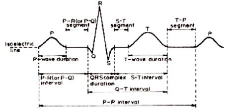
Figure 1. ECG Signal
An effective as well as efficient analysis of ECG signal depends upon accurate and reliable detection of the P, Q, R, S and T waves [32]. The detection of QRS complex is the most important task in automated ECG signal analysis because once the QRS complex is being identified, a more meticulous assessment of ECG signal is doable by including the heart rate and ST segment [5-6]. Number of algorithms developed have been discussed in the current paper for early detection as well as classification of these ECG signals.
Many algorithms have been developed for detection, feature extraction as well as classification of the ECG signals. Ramli and Ahmad [7] have used a cross correlation analysis technique for extracting the vital features from 12 lead ECG signals. Using the cross correlation techniques the values being identified can be used to predict the type of arrhythmias.
Tadejko and Rakowski [8], introduced an automatic classifier with K-self-organizing maps (SOM) and LVQ algorithms. This paper compares the QRS complexes for classification and then preprocesses the ECG morphology features. Various types of arrhythmias related to cardiac is followed in Table 1. The performance of algorithms is to examine the beats either in the form of normal or arrhythmia condition.
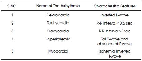
Table 1. Various Abnormalities and their Characteristic Features
Xu and Liu in [9] proposed the SVM algorithm for ECG QRS complex detection as well as the RR interval determination. They have used the Slope vectors for the extraction of features of ECG wave and also the nonlinear amplification have been used to improve S/N. This paper introduces high accuracy and fast response to find the QRS detection.
Manpreet Kaur and Arora [10] anticipated the K- means clustering along with Squared Euclidean distance for the ECG signals analysis. For ECG feature extraction, the parameters identified as wave shape, duration and amplitude. Using K-means clustering technique, clustered K is summed and minimized to a centroid distance.
F. De Chazal and Reilly [23] determines the PVC from normal beats. For feature extraction of ECG signals, the combination of morphological based features and time interval based features have been proposed. For the classification of ECG signal, the MLP with different number of hidden layers and algorithm according to, Radial Basis Function Neural Network (RBFNN) and Probabilistic Neural Network (PNN) is used. The simulation results show that about 97.14% for classification is achieved [15]. For simulation purpose, the MIT-BIH arrhythmia database is used.
S. Mitra et.al [11] determines a rough-set theory for the analysis of ECG signal. A rule-based rough-set decision system is developed from time-domain features to make an inference engine for arrhythmia detection. This technique helps to optimize rules for cardiac- arrhythmia detection, by which the complexity of Neural Networks (NN) can be omitted [16]. Currently, the system is tested with three types of ECG datasets as Normal, Myocardial Ischemia and Myocardial Infarction and the accuracy of all these types is obtained for both the trained and untrained dataset.
Castro et al. [12] anticipated a wavelet transform algorithm for feature extraction of an ECG signal and identification of abnormal heartbeats. The ECG signal is first denoised by a soft or hard threshold and then each cycle is simplified into a coefficient vectors using the Optimal Wavelet Function (OWF). The analyzed ECG signal coefficients are divided into P, Q, R, S and T to obtain a feature vector of the signal cycles.
Nazmy et al. [13] determines the ANFIS algorithm for classification of an ECG wave. The ECG feature extraction is done using the Independent Component Analysis (ICA). The power spectrum and input is being provided by the RR interval of ECG signal.
Alan and Nikola in [14] have introduced Chaos theory for feature extraction of ECG signals. Various methods comes under the Chaos theory, including correlation dimension, phase space and attractors, central tendency measure, spatial filling index, and the approximate entropy are also determined.
Yuksel and Bekir [17] have represented ANN to classify the ECG arrhythmias.
Zhu et.al, [18] determines the application of ANN for ECG abnormality detection. In this paper, to analyze the performance, the SOM network, BP and LVQ network were used and the overall accuracy of these networks has been acheived. [19,[20],[21]] also presented a comparison of how the neural networks classify the patterns from training data and recognizes if testing data holds that patterns of ECG signal.
El-Khafif et al. [19] anticipted ANN model to make a diagnosis of the ischemic heart disease from normal ECG signals.
Hosseini et al. [22] have anticipated the use of a twostage Feed Forward Neural Network (FF-NN) for ECG signal classification in which, they have elected two network architectures on the basis of one stage and two stage Feed Forward Neural Networks (FF-NN) to recognize heart arrhythmias.
Manimegalai et al. [28] have determined the use a discrete wavelet transform based system for detection and feature extraction of P, Q, R, S and T waves and in which, they found that this technique consumes less computational time and has better accuracy for ECG classification, analysis and characterization of normal and abnormalities of ECG. In [24,25,[26],27], the neurofuzzy technique has been proposed to configure the experimental data.
Golpayegani and Jafari [29] proposed a comparative evaluation of recital of ANFF with MLP neural networks and it has been found that, the time of training required for the ANFF was much shorter than time required by MLP.
Owis et al. [30] have determined the correlation dimension and largest Lyapunov exponent parameters to sculpt the chaotic nature of different classes of ECG signals. The anticipated implementations were used to compute these features belong to five different types of ECG signal taken from the MIT-BIH Arrhythmia Database.
The introduction of neural network was done in 1943 by the famous neurophysiologist, Warren McCulloch and logician, Walter Pits. ANNs are biologically inspired networks which has many applications in the fields such as, pattern recognition, classification, etc. Typically, multilayer feed forward neural networks can be trained as non-linear classifiers using the generalized Back Propagation Algorithm (BPA) [31-33]. The back propagation algorithm is a type of supervised learning algorithm, in which a Mean Square Error (MSE) function is defined, and the basic aim of the learning process is to reduce the overall system error to a minimum.
The technique of classification is done by logical and accurate neural network algorithm called Back Propagation Network (BPN). The learning algorithm of the multilayer perception requires a differentiable activation function, but frequently used is the logical function (nonlinear, monotonic, increasing, differentiable). The term back propagation states the backward propagation of an error signal through the network. After propagating a pattern through the feed forward network, the output pattern is compared with a given target and the error of each output unit is calculated. The error of each output unit is propagated backwards to the input layer - back propagation. Finally, the errors of the unit are used to modify the weights.
The Back-Propagation Neural Network (BPNN) usually allows practical attainment of each input/output mapping information within each multilayer networks. Basically, BPNN executes the algorithm of gradient descent search to minimize the Mean Square Error (MSE) between the preferred output and the actual output of the network by adjusting their weights. Back propagation (BPNN) algorithm is highly in use because in most classification problems, the reason is the characteristics of the generalized data rule [33,34].
It is a forward multi-layer network, which trains the network by using the error back-propagation algorithm. The Back Propagation Network algorithm (BPN) was proposed by Rumelhart et al. in 1986, because of its simple structure, multiadjustable parameters, much training algorithm and good operational performance, by which this network has got a wide range of practical application. The network structure of the three-layer BP neural network is shown in Figure 2.
Figure 2. Structure of ANN
The structure of ANN shows that, this neural network consists of an input layer, a middle layer (hidden layer) and an output layer. In this, there is a complete connectivity between the upper and lower layers and there are no connections between the neurons in each layer. The input layer of the signal needs to spread towards the hidden layer nodes and is transformed by the function and then the transmission of input signal of the hidden layer nodes to the output layer nodes. Typically the transfer function of BP neural network is Sigmoid Type differentiable function, which can attain random nonlinear mapping between the input and output, from this result, this type of network has been used in wide applications of Pattern Recognition, Function Approximation and other areas [11] [35-36] .
Wavelet transform is a periodic scale representation that has been utilized effectively as a part of wide scope of utilizations, specifically signal compression. As of late, Wavelets have been connected to few issues in Electro cardiology, including data compression, examination of ventricular late possibilities, and location of ECG characteristic points. The wavelet transform is a straight operation that breaks down the signal into various scales identified with recurrence parts and examines every scale with a specific determination. The WT utilizes a brief span interim for assessing higher frequencies and while interim for lower frequencies. Because of this property, high frequency components of short term can be watched effectively by Wavelet Transform. One of the benefits of the Wavelet Transform is that, it can break down signs at different resolutions, which permits precise component extraction from non-stationary signs like ECG. A group of breaking down wavelets in the time recurrence area is acquired by applying a scaling component and an interpretation element to the fundamental mother wavelet. Figures 3 and 4 show the levels of ECG decomposition.

Figure 3. Levels of ECG Decompostion
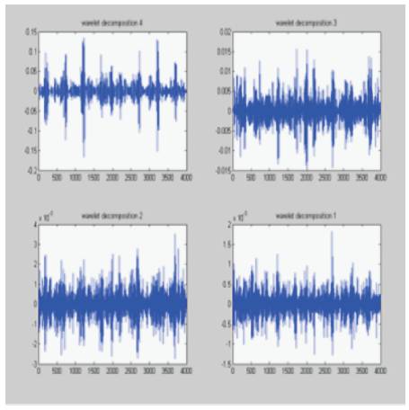
Figure 4. Levels of ECG Decomposition
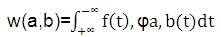
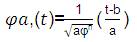
Wavelet change of a sign f(t) is characterized as the entirety of over unsurpassed sign duplicated by scaled, moved forms of the wavelet capacity ψ, which is given by,
The DWT is adequate for most of the applications and for the recreation of the sign. The DWT gives enough data, and it gives a huge decrease in the calculation time. Here, the authors have discrete capacity f (n) and the meaning of DWT is given by:
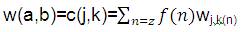
where,
 is a discrete wavelet transform,
In the DWT examinations, the signal at various frequency bands and resolutions is deteriorated into a 'coarse estimation' and 'point by point data'. Two arrangements of capacities are utilized by DWT, the scaling capacities (connected with the low pass channel) and the wavelet capacities (connected with the high pass channel). The sign is shifted by going it through progressive high pass and low pass channels to acquire variants of the sign in various recurrence groups. The essential thought behind wavelets is to dissect as per scale. These are capacities that fulfill certain numerical necessities and are utilized as a part of speaking information on different strategies. Calculation process information at various scales or resolutions. In the event that the sign with a substantial window, would see gross elements. Additionally, signal with a small window have a small element.
At one point, the order is finished by Back Propagation Network. The learning calculation multilayer discernment requires a differentiable actuation capacity, much of the time is logistic capacity utilized (non-direct, monotonic, expanding, differentiable) [15]. The term back proliferation implies the regressive spread of a blunder signal through the system. Subsequent proliferating an example of the system - encourage forward, that the yield example is contrasted and a given target and the mistake of every yield unit is ascertained [16]. This blunder is proliferated in reverse to the data layer - back spread. At long last, the blunders of the units are utilized to adjust the weights.
The data are collected from MIT-BIH database. In this paper, 34 signals are used for training and from this, 15 signals are taken for testing.
These techniques accomplishes the separation in the normal and abnormal heart rate, which are analyzed by back propagation neural system. Table 2 explains the outcomes of the analysis done. This technique demonstrates the ongoing application execution parameter that can precisely judge the exactness.
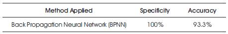
Table 2. Performance Parameters
ECG signals required for investigation are gathered from Physionet MIT-BIH arrhythmia database. The database contains 48 records, each containing two-channel ECG signals for 30 min length of time chosen from 24-hr recordings of 47 distinct people. The techniques were produced under MATLAB. Subsequent to the utilization wavelet, change in electro cardiology is generally a new field of examination, numerous methodological perspectives (Choice of the mother wavelet, estimations of the scale parameters) of the wavelet strategy that will require further examinations with a specific end goal to enhance the clinical helpfulness of this novel sign handling framework. Simultaneously; analytic and prognostic noteworthiness of wavelet strategies in different fields of electro cardiology should be set up in vast clinical studies. Extremely straightforward and quick dependable techniques are proposed in this paper. This system is anything but difficult to perform and it need not bother with any complex scientific estimations, for example, Fourier strategies, and cross-connection. The ANN classifier was nourished by three parameters, spectral entropy, Poincare plot geometry and biggest Lyapunov type (LLE) obtained from the heart rate signs that were analyzed.