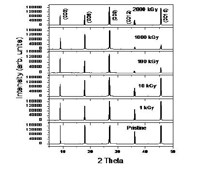
Figure 1. X- ray diffraction pattern for pristine and gamma irradiated muscovite mica.
Present work reports the structural and optical properties of pristine and gamma irradiated muscovite mica. The XRD spectra are used to estimate structural parameters such as crystallite size and micro strain of pristine and irradiated samples. Williamson Hall analysis is employed to calculate the crystallite size and micro strain of pristine and irradiated sheets. UV-VIS analysis provides the value of optical indirect, direct band gap and Urbach energy. It was found that the value of optical indirect and direct band gap increases with the increase of gamma dose upto 100 kGy and then decreases with further increase in gamma dose upto 2000 kGy. Thus, the increase of optical band gap makes natural muscovite fits for efficient optoelectronic devices.
Muscovite mica [KAl2 (AlSi3O10) (F, OH)2] is an abundant mineral on the earth and has a layered structure of aluminium silicate sheets weekly bonded together by layers of potassium ions. These potassium ion layers produce the perfect cleavage [1]. Muscovite is used chiefly as an insulating material and for fireproofing and are presently being used as dosimeter for monitoring absorbed doses in radiation rich environments. It is well established that the exposure of any material to ionizing radiations produces changes in the microstructural properties of the material, which in turn affects the structural and optical properties [2]. Thus, it is important to understand the effect of ionizing radiation on the structural and optical properties of these materials.
A lot of information is available on the optical [3-7] properties of natural muscovite mica without irradiation. Moreover, the effect of gamma irradiation on these properties has not been investigated so far. Therefore, in the present work, the effect of gamma dose on structural and optical properties of natural muscovite mica have been investigated to utilize this material for innovative applications in radiation technology and opto-electronic devices.
Thin sheets of natural muscovite mica (procured from Bhilwara mines in Rajasthan) samples were irradiated with 60Co source to different doses ranging from 1 kGy to 2000 kGy at IUAC, New Delhi. The structural changes were studied using XRD-7000 SHIMADZU X-ray diffractometer. The optical changes were analysed with UV-1800 Shimadzu UVVis spectroscopy in the wavelength range 200-600 nm.
Figure. 1 shows the XRD pattern for pristine and gamma irradiated natural muscovite mica. Five peaks are observed in pristine muscovite mica at 2θ = 8.99o , 17.90o ,26.94o , 36.12o and 45.59o corresponding to crystallographic planes (003), (006), (009), (0012) and (0015), respectively. No shift in peak positions is observed after gamma irradiation.
Figure 1. X- ray diffraction pattern for pristine and gamma irradiated muscovite mica.
The average particle size and the microstrain for the pristine and gamma irradiated samples were determined using the Williamson Hall (W-H) analysis [8]. Figure. 2 shows the W-H plots for the pristine and the gamma irradiated muscovite at different absorbed doses. The values of crystallite size (D) and microstrain (e) obtained from the intercept and slope of WH plots are listed in Table 1. The crystallite size increases with the gamma dose upto 100 kGy; microstrain decreases in the dose range of 1- 100 kGy. For gamma doses upto 2000 kGy, the crystallite size decreases, however, unlike the microstrain which increases. This means that in the lower gamma dose range, 1 -100 kGy, the irradiation binds the crystallites in clusters, increasing their size and decreasing the microstrain. At higher gamma doses, i.e. 1000, 2000 kGy, the crystallite size decreases and the microstrain increases which indicates that the muscovite moves to a disordered structure [9].
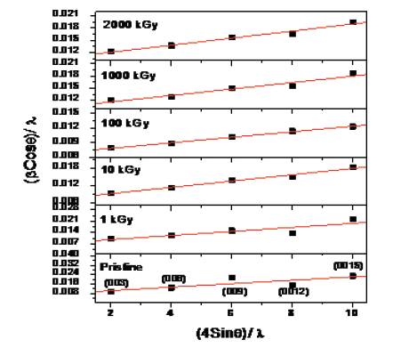
Figure 2. W-H plots for the pristine and gamma irradiated muscovite mica.
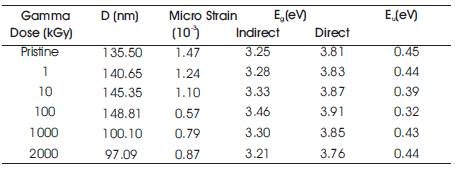
Table 1. Variation of structural and optical parameters with gamma dose
Figure. 3 presents the UV–visible absorption spectra of the pristine and gamma-irradiated (1-2000 kGy dose) natural muscovite mica samples. The absorption edge is shifted towards lower wavelength values by increasing the dose up to 100 kGy. However, by further increasing the dose upto 2000 kGy, the absorption edge shifts towards higher wavelength values.
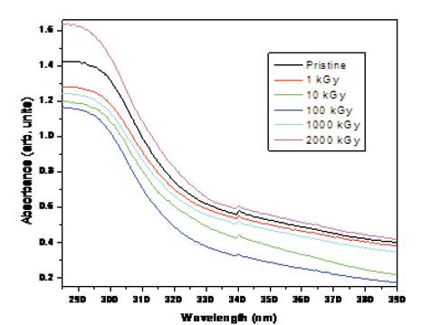
Figure 3. UV-Vis spectra of pristine and gamma irradiated muscovite mica.
The optical band gap was determined using the following equation [10]:
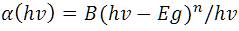
where hv is the energy of the incident photons, Eg is the value of the optical energy gap between the valance band and the conduction band, B is a constant. The value of n as 1/2 and 2 stands for direct allowed and indirect allowed transitions, respectively. The calculated values of direct and indirect optical band gap for pristine and irradiated samples are given in Table 1. The values of optical indirect and direct band gap increase from 1 to 100 kGy and decrease by further increasing the gamma dose upto 2000 kGy.
The irregularities in the band gap level can be defined in terms of Urbach energy (Eu) and were estimated using the relation [11]:
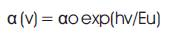
where αo is a constant, Eu is an Urbach energy and v is the frequency of radiation. The calculated values of Urbach energy for pristine and gamma irradiated muscovite at different doses are listed in Table 1. The decrease of the Urbach energy at increasing γ-doses, from 1 kGy to 100 kGy, indicates the decrease in the structural disorder. By further increasing the dose upto 2000 kGy, the Urbach energy increase may be attributed to increasing of the structural disorder.
Gamma irradiation of upto 100 kGy leads to the increase in crystallite size and reduction in the defects, structural disorder and microstrain. Thus, irradiation of natural muscovite mica with gamma rays improves its crystallinity. The results of UV-VIS spectra show that the direct and indirect optical band gap can be monitored with the help of gamma irradiations. The increase of optical band gap with gamma irradiation makes natural muscovite fit for efficient optoelectronic devices.
Authors are highly thankful to Health physics and material science group at IUAC, New Delhi for providing the Gamma Irradiation, XRD and UV-Vis facilities. Financial support given by UGC, New Delhi, in the form of Senior Research Fellowship is also gratefully acknowledged.