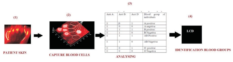
Figure 1. Typical Block Diagram for Digital Image Processing and Analysis
The primary objective of this work is image processing based blood groups identification that utilizes the digital image matching process without using needles. Now-a-days, the blood group testing uses needles and also utilizes some chemicals, optical plates, cotton cloths, etc. Above biomaterial disposal in the environment is very dangerous and also creates environmental soil pollution. Non-bio degradable materials are the main pollution sources in soil. Technology and various researches have dominated to save human blood and to control the soil pollution and thus the present situation is met. The novel blood group testing is done by finding blood group of a patient without piercing the skin. This article explains a method to determine the human blood type by applying digital image processing to understand the image of artificial vessels underlying the skin. The research includes Multicore wavelength light sprinkling method, where light passes through the vessels for classifying the blood cells based on exact antigens on the red blood cell surface. The transferrable camera along with photo-detectors forms the basic detector structure and is used to detect the light distribution produced by blood cell to determine the blood type. This research presents a current state-of-the-art in optimizing digital image processing based blood group identification, which provides a clear vision of the latest top research advances in image processing with the help of MatLab and Embedded C program.
The blood group testing is an art of blood transaction. America, Germany and other countries use various solutions for analyzing blood groups as a dynamic factor (Berlitz et al., 2011). Man by nature has any one of the Blood groups, such as A, B, AB, and O (Selvakumari, 2011). The blood groups “AB” is called the “Universal acceptor” and the people with “O” group are called “Universal donor ” (Geisow & Beaven, 1997). During blood transfusion, any incompatibility may even lead to the decease of a person. Hence it is utmost important for every person to identify their blood group. In general, the specialists in laboratories are seminal about the blood group by using ABO blood analysis method. This analyze is under the basis of aggregation method of antigen and antibody (Blood Type, n.d.). Now the antibody is a collection of toxins, bacteria, foreign blood cells, and the cells of resettled organ. Using physical analysis, one can get better results, but in handling with wide range of samples, the examining person experiences a very tough job and may make mistakes in maintaining the blood samples and records accurately (James & Wickerhauser, 1972). In such situation, here a new resolution is proposed to diagnose the blood group (Hearnshaw, 2004). In this new technique, the optical source and photo-detector are used to analyse the ABO blood grouping (A, B, O) and Rhesus type testing (Rh +ve and -ve). Currently, a mediocre of 300 to 500 blood samples are analyzed within 3 to 4 hours which can be carried out only by specialists, but when the proposed device is used, the same mission can be accomplished within a very short period of time with correct precision. One more added improvement of this proposed scheme is that it is inexpensive. Before a blood transfusion, it is essential to RESEARCH PAPERS perform certain tests (Uda et al., 1977). One of these tests is the fortitude of blood type and this test is essential for the apprehension of a safe blood transfusion, so as to manage a blood type that is compatible with the type of receiver (Fathima, 2013). There is a certain emergency condition when a patient’s life is at risk, it is necessary to administer blood immediately (Schwyzer & Hill,1977a). The tests currently available require moving the test center, which may not determine the blood type better and as the precise blood type ‘O’ negative is considered as a general donor, it provides less risk (Ferraz, 2013). However, despite the risk of minimum incompatibilities that sometimes occur during transfusion can cause decease of the patient and it is essential to avoid them, when directing blood based on the principle of universal donor only (Romaniuk & Gajda, 2013). Thus, the ideal situation determined would be administering the blood type of the patient even in reserve situations, ie., compatible blood type from the first unit of blood transfusion (Brinkhues et al., 1992). Moreover, the pretransfusion tests are performed physically by technician's analysts, which sometimes lead to the manifestation of human errors in procedures, reading, and deducing of results (Hosoi, 2008). Since these human errors can translate on fatal concerns for the patient, being one of the most substantial causes of incurable blood transfusions is extremely important to systematize the procedure of these tests by performing reading and elucidation of the results (Satoh & Itoh, 2006). Blood group is collection of blood based on the manifestation or absence of congenital antigens (Schwyzer & Hill, 1977b).
During digital image processing, the blood group is essentially identified as an Embedded C program. The optical sensor directly passes the photons in to the patient’s skin. The photon captures the blood cells (red blood cell, white blood cell) after which the photons are reflected back to the photo cell detector. The photo cell detector embeds with Embedded C program as shown in Figure 1.
Figure 1. Typical Block Diagram for Digital Image Processing and Analysis
The program contains various loop structures. Embedded C program loop concepts contain large types of looping program. For convenience, the switch case method is choosen and the switch case method contains various blood group cases (A+ , A1+, A-, AB+, O-, etc…). The switch case processes before matching the red blood cell in all already and physically captured ABO blood group flashes (manual research laboratory) including laser passing process as shown in Figure 2.
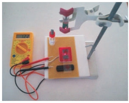
Figure 2. Physical Collection of ABO Blood Group Flashes
The first step is matching the image processing flashes using manual research laboratory images. Image processing matches the images one by one, where the image that matches with the matching image denotes the blood group name (eg. A1+). Finally blood group type is displayed in 20 x 4 Liquid Crystal Display (LCD) as shown in Figure 3 after doctor or lab technician has certified it to the patient.
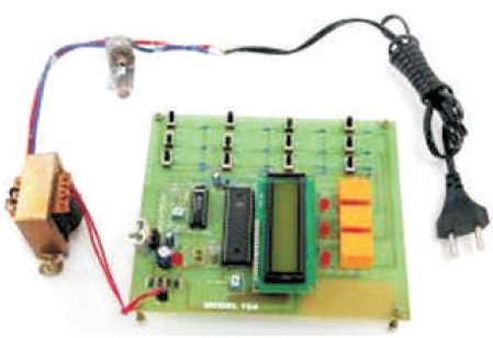
Figure 3. Proposed Digital Image Processing
The ABO blood collection arrangement involves two antigens and two antibodies that originate in human blood. The two antigens are antigen A and antigen B. The two antibodies are antibody A and antibody B. The antigens are present on the red blood cells and the antibodies in the serum concerning the antigen property of the blood. All human beings can be divided off the record into 4 groups, those with antigen A (group A), those with antigen B (group B), those with both antigen A and B (group AB), and those with neither antigen (group O). The antibodies present together with the antigens are found. First one is Antigen A with antibody B, second one is Antigen B with antibody A, Antigen AB has no antibodies, Antigen nil (group O) with antibody A and B. There is an agglutination reaction between similar antigen and antibody (For example, antigen A agglutinates the antibody A and antigen B agglutinates the antibody B). Thus, transfusion can be considered safe as long as the serum of the beneficiary does not contain antibodies for the blood cell antigens of the donor (Blood Type, n.d.). The ABO system is the most significant blood-group system in human-blood transfusion. The associated anti-A and anti- B antibodies are usually immunoglobulin M, abridged IgM, antibodies. ABO IgM antibodies are created in the first years of life by sensitization to environmental substance, such as food, bacteria, and viruses. The original terminology used by Karl Landsteiner in 1902 for the classification was A/B/C; in later publication "C" became "O". "O" is often called 0 in other language as shown in Table 1(Blood Type, n.d.).
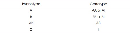
Table 1. ABO Blood Group System
An optimizing blood group determination analysis proposal is based upon normal blood group determination that replaces the digital image processing based blood group identification (Paulson et al., 1977). This novel blood group determination device consists of a optical sensor (primary source), photo- cell detector, embedded C program with 20 x 4 LCD display. Net multicore wavelength reflection processes after producing the image pattern. After the image pattern fed by the photo cell detector, there is also a capture of blood cells, but we want only for digital outputs so we are using Embedded C program with switch case methods so scanning image matches the already programmed images, which one of the matching flashes is donated by the blood group name. Finally the blood group name fed by the 20 x 4 LCD display is shown in Figure 3.
The results which are obtained from the work done are discussed. The digital image processing based blood group analysis shows that the output efficiency is compared in normal laboratory based blood group analysis and its output efficiency are shown in Figures 4 and 5. The normal laboratory based analysis efficiency is very low, more time consuming and % efficiency is also low. The digital image processing analysis output efficiency is linearly increased as shown in Figure 5 and consumes less amount of time, and efficiency is also high.
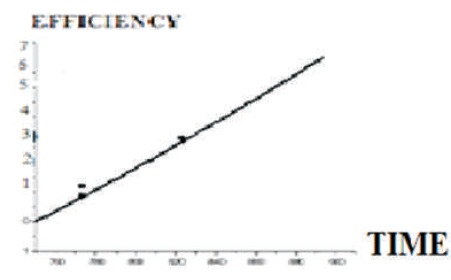
Figure 4. Digital Image Processing Based Blood Group Analysis
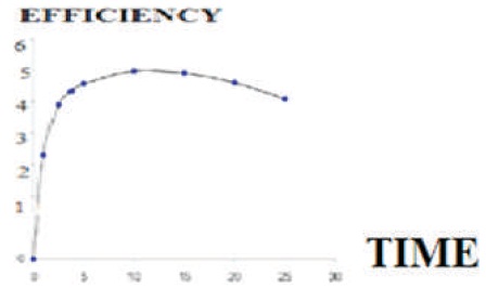
Figure 5. Normal Laboratory Based Blood Group Analysis
This paper presents a digital image processing based blood group analysis, which is based on the concept of Embedded C program. This aids the present biomedical research demand and paves a way to reduce the time requirement and efficiency improvement. However, to tune the gain of image processing and programming process is a challenging task. Thus this proposed method developed ultimately reduces the blood group analyzing steps requirement and have generated a cost-effective method. This method also explains a novel system that brings this development of technology much closer to marketable practicality, where the prime motive of the digital image processing based blood group analysis not to create any form of toxic waste, other pollutants and also reduce soil pollution. This aids the people to safeguard the valuable human life during emergency blood transfusion. This user-friendly modified blood group analysis will find applications for patients residing in remote hill stations and physically challenged patients.