Image segmentation is one of the most widespread means to classify the pixels of an image correctly in decision oriented applications. Image segmentation is a technique that partitions an image into uniform and non overlapping regions based on some likeness measure [1]. The major role of segmentation is in the field of biomedical applications. This paper compares fuzzy cmeans segmentation method with region growing method in the presence of noise level. Fuzzy C- means is a method of clustering which allows one pixel to belong to one or more clusters based on some given criteria. Region growing is a procedure that looks for group of pixels with similar intensities. It starts with a pixel or a group of pixels called seeds that belong to the structure of interest. In order to evaluate the performance of segmentation methods, parameter accuracy is used for estimation. Segmentation results will be more accurate if medical images are noise free. Therefore, it is necessary to use denoising technique before segmentation. Denoising of images can be done either by using spatial domain [2] or frequency domain [3]. Denoising in spatial domain reduces the noise but creates blur on the images [4] . In order to achieve both noise reduction and feature preservation, wavelet-based methods are of particular interest. In the wavelet domain, the noise is uniformly spread throughout coefficients while most of the image information is concentrated in a few large ones [5]. Therefore, the first wavelet-based denoising methods were based on thresholding of detail sub band coefficients [6] followed by inverse wavelet transform which yields denoised image. Most of the work on wavelet based denoising technique has been carried out by using orthogonal wavelet transform [2,7] . But the limitations of orthogonal wavelet is that, it is not invertible. Designing orthogonal wavelet does not allow degree of freedom i.e. no possibility to construct symmetric wavelet function. Therefore, the authors proposed a new method of image denoising as Biortogonal wavelet transform. Biorthogonal wavelet denoising technique is used to remove the noise present in the image while preserving the image characteristics, regardless of its frequency content which increases the robustness of the segmentation approach against noise and has been demonstrated with the aid of experimental results.
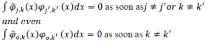
Biorthogonal wavelets exhibit the property of linear phase which is needed for signal and image construction. Two wavelets  and
and  are used. function is used for analysis and the other function is used for synthesis [4]. Also, two scaling functions and are used to produce different multiresolution analysis. The scaling function , and wavelet function , are related by duality in the following sense:
Step 1: Choose biorthogonal (bior 1.3) wavelet transform and decomposition level as three.
Step 2: Apply hard thresholding method with sparsity parameter (a>1).
Step 3: Apply inverse wavelet transform to reconstruct the denoised image.
This section deals with two segmentation approach region growing method and fuzzy c-means method for the effective segmentation of noisy medical images.
Region growing is a procedure that looks for groups of pixels with similar intensities. It starts with a pixel or a group of pixels called seeds that belong to the structure of interest. Subsequently, the neighboring pixels with the same properties as seeds are appended gradually to the growing region until no more pixels can be added. The object is then represented by all pixels that have been accepted during the growing procedure. The advantage of region growing is that, it is capable of correctly segmenting regions that have the same properties and are spatially separated, and also it generates connected regions [8], [9].
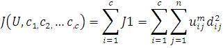
Fuzzy C- means is a method of clustering which allows one pixel to belong to one or more clusters. The cluster patterns are classified in such a way that samples of the same group are more similar to one another than samples belonging to different groups [10]. Fuzzy c-means algorithm is based on minimization of the following objective function.
(2)
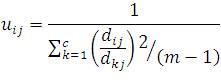
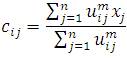
uij is between 0 and 1; C is the centroids of cluster I; dij is the Euclidian distance between the ith centroids and jth data points; mε[1,∞] is a weighting function. Fuzzy portioning of known data samples is carried out through an iterative optimization of the objective function,
This iteration will stop when 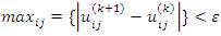 where,∈ is a termination criterion between 0 and 1, whereas k represents the iteration steps. This procedure converges to a local minimum or a saddle point of jm
3. Experimental Results and Performance Evaluation
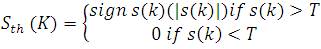
The proposed algorithm have been implemented using three stages-pre-processing, denoising, and segmentation. For denoising, biorthogonal wavelet transform via hard thresholding is used. Hard thresholding is described as a process of setting the elements to zero whose absolute values are lower than the threshold and is defined as
where T is the threshold.
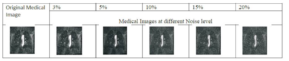
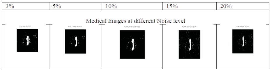
Experiments were conducted on MRA Cardio images. These images were obtained from Physionet.org [11]. These images are used for comparison between fuzzy cmeans and region growing method for different additive white Gaussian noise level as 3%, 5%, 10%, 15% and 20%. The quality of the segmentation results is calculated in terms of segmentation accuracy.
3.1 Segmentation Accuracy (SA)
Segmentation accuracy determines the eventual success or failure of computerized analysis procedures, and for this reason a considerable care is taken to improve the probability of accurate segmentation [12].
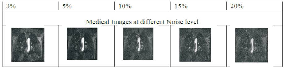
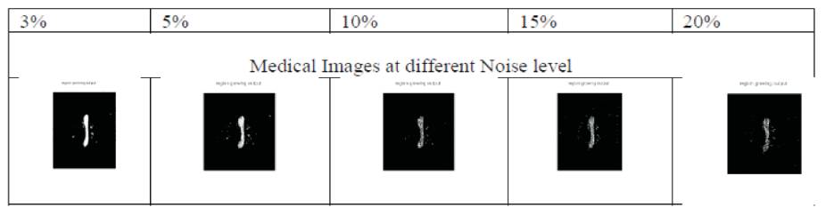
The experiment was conducted for more than 75 different medical resonance angiography of cardio images in presence of noise. This paper discussed only one sample (E1154S71000.png) with additive White Gaussian noise at different noise level .The performance analysis of above discussed segmentation approach without denoising is shown in Figure 1. In order to evaluate the robustness of the segmentation approach with denoising,a biorthogonal wavelet transforms using hard thresholding is used. The additive white Gaussian noise at different level and corresponding denoised output at different level are shown in Table 1 and 2 respectively. Also, segmentation results using region growing method and fuzzy c- means for different noise level are shown in Tables 3 and 4. From Table 3, it is also observed that, if noise level increases above 5% than the segmentation results, deteriorate for region growing method. Table 4 shows that, if noise level increases above 15% than segmentation results, deteriorate for fuzzy c- means method.
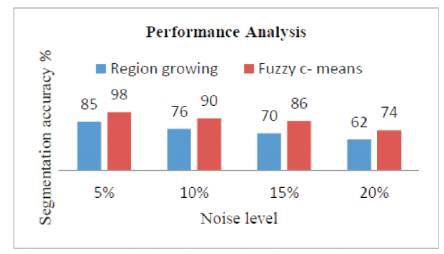
Figure 1. Segmentation Accuracy of Fuzzy c-means and Region Growing Method without Denoising MR Images with different Noise Level
Conclusion
In this paper, region growing and fuzzy c means methods are used for medical resonance angiography for different noise level at 3% 5%, 10%, 15% and 20%. All medical images are prone to noise either at the time of transmission or at the time of acquisition. So, denoising is the primary step before applying segmentation. The authors used biortogonal wavelet transform denoising technique to improve the performance of segmentation results. The performance of algorithms is measured using Segmentation Accuracy (SA) and the results shows that the fuzzy c- means provides good segmentation result as compared to region growing method in the presence of noise, but fuzzy c- means segmentation result, deteriorates the noise level increase after 15%. To overcome this, noise is the main topic of our future work.
References
[1]. Dong, G. and XieM. (2005). “Color clustering and learning for image segmentation based on neural networks”, IEEE Transaction on Neural Networks, Vol.16, pp. 925-936.
[2]. Khare, A., and Tiwary,U. S.(2005). “Soft- Thresholding for denoising of medical images A Multiresolution Approach”, International Journal of Wavelets, Multi resolution and Information Processing, Vol. 3, pp. 477–496.
[3]. Egiazarian,K., and Astola,J. (1999).“New Algorithm for Removing of Mixed (White and Impulsive) Noise from Images”, Nonlinear Image Processing X, Proceedings of SPIE, Vol. 36, No. 46, pp. 78-89.
[4]. Prakash,O., Khare,A., and Khare,M. (2013). “Image Denoising technique based on soft thrsholding of boiorthogonal wavelet coefficients ”, National conference on Advances in Mobile Communication, Networking and Computing, pp. 155-159.
[5]. Forouzanfar,M., Abrishami-MoghaddamH., and GhadimiS (2008). “Locally adaptive multiscale Bayesian method for image denoising based on bivariate normal inverse Gaussian distributions,” International Journal of Wavelets, Multiresolution and Information Processing, Vol. 6, pp. 653-664.
[6]. Mallat, S.(1998). “A Wavelet Tour of Signals Processing”, Academic Press, London
[7]. Binh, N.T., and Khare,A. (2010). “Multilevel threshold based image denoising in curvelet domain”, Journal of Computer Science and Technology, Vol. 25, pp. 632-640.
[8]. Hanmandlu ,M., Dubey, R.B., and Gupta, S.K. (2010). The Brain MR Image Segmentation Techniques and Use of Diagnostic Packages, Academic Radiology, Vol.17, pp. 658-671.
[9]. Ohya, J., Luo, L. and Xu, R.( 2012). “ Segmentation of Brain MRI”, Advances in Brain Imaging, Vol. 8, pp.143-170.
[10]. Zheng,L., Zhou,J., and Wen,P. (2007). "Spatial Credibility Clustering Algorithm in Noise Image Segmentation", IEEE International Conference on Industrial Engineering and Engineering Management, pp. 543 -547.
[11]. www.Physionet.org.
[12]. Ahmed,M.N., Yamany,S.M., and Mohamed,N. (2002). “A Modified Fuzzy c-means Algorithm for Bias field estimation and segmentation of MRI data”, IEEE