Abstract
Image fusion is the process of producing a single image from a set of input images . The fused image should have more complete information which is more useful for human or machine perception. The fusion of images is an important technique within many desperate fields such as remote sensing, robotics, manufacturing , intelligent Systems, medical applications etc. With the evolution of imaging technology, an increasing number of image modalities becomes available such as CT, SPECT, PET, MRI etc. Each modality has its strength and weakness. For example, SPECT would be able to show image functional behavior of organs, but has low resolution with diffuse boundaries. Because of this it is difficult to identify specific organs or tissues. On the other hand, X-ray Computed Tomography (CT) and MRI provide images with high resolution and sharp boundary information. To preserve all the complementary informations provided by different modalities in a single image, image fusion is performed, which is useful for human visual and machine perception and for better data interpretation.
An intensity based fusion method is developed using JAVA for fusing multimodal medical images. This fusion method is applicable for fusing images that is obtained from two different modalities (Computer Tomograhy- CT and magnetic resonance imaging (MRI). This image fusion software reads two images and combines them into a single highly informative image with maximum content. The program is tested using CT-MRI images that is fused using methods like wavelet pyramid ,fast point based fusion, using MATLAB platform etc.
Introduction
Image fusion is the process of combining relevant information in two or more images of a scene into a single highly informative image. Relevant information depends on the application under consideration. The main advantages of Image Fusion are improved reliability (by redundant information)and improved capability (by complementary information). The Objectives are to extract all the useful information from the source images, and not introducing artifacts or inconsistencies which will distract human observers, or the following processing and reliable and robust imperfections such as mis-registration. Modern Medical technology provides a wide range of scanning and measurement systems. Radiologists and surgeons often have to interpret a large number of images from different types of scanners. To support diagnosis or to manage a patient's therapy, there is an increasing need for image fusion, i.e. to relate and extract information from medical images of the same patient acquired with different techniques. The data sets provided by the latest imaging techniques are confusing to interpret for the physicians .The images are to be presented to the physicians in such a way, that they can be easily and correctly interpreted without any loss of information. The aim of the study is to develop an effective registration and fusion method to improve accuracy and efficiency in diagnosis and planning of therapy.
Overview of Medical Multimodalities
Modality -CT and MRI provide anatomic (structural) detail and can help identify abnormal masses or distortion of normal structures by disease. Fusion of CT-MRI has been described in the paper [7] using multiresolution analysis method. We have found a varied list of applications ,and is summarized in the following [10,11,12]. Fusion of PET-MRI has been described in variety of applications[7,10].
CT-SPECT - SPECT imaging is the image modality which has the highest accuracy in detecting bone inflammation. The fusion of these two modalities has been done in several applications[10,13].The registration of MRI-US modalities has been done in various applications[10].
Object - The Object in medical image registration is the part of the anatomy involved Brain/Head. The large part of the literature on image registration concerns head or brain images[7,10,12]. Liver Image registration from the two modalities, CT and SPECT, acquires both structural and functional information, and therefore is very useful for clinical diagnoses. The registration and fusion of CT and SPECT images are studied [13].
Image fusion Methodology
Evolution
Simple image fusion attempts - The primitive fusion schemes perform the fusion right on the source images, which often have serious side effects such as reducing the contrast.
Pyramid-decomposition based image fusion methods- With the introduction of pyramid transform in mid-80's, some sophisticated approaches began to emerge. People found that it would be better to perform the fusion in the transform domain. Pyramid transform appears to be very useful for this purpose. The basic idea is to construct the pyramid transform of the fused image from the pyramid transforms of the source images, then the fused image is obtained by taking inverse pyramid transform. Here are some major advantages of pyramid transform: It can provide information on the sharp contrast changes, and human visual system is especially sensitive to these sharp contrast changes and it also provides both spatial and frequency domain localization. Several types of pyramid decomposition are used or developed for image fusion, such as: Laplacian Pyramid ,Ratio-of-low-pass Pyramid, Gradient Pyramid etc.
Wavelet based decomposition method- Image fusion began to receive increasing attention, more recently, with the development of wavelet theory. People began to apply wavelet multiscale decomposition to take the place of pyramid decomposition for image fusion. Actually, wavelet transform can be taken as one special type of pyramid decompositions. It retains most of the advantages for image fusion but has much more complete theoretical support.
With the evolution of imaging technology, an increasing number of image modalities become available such as CT, SPECT, PET, MRI etc. Each modality has its strengths and weaknesses. For example, SPECT would be able to show image functional behavior of organs, but has low resolution with diffuse boundaries. Because of this, it is difficult to identify specific organs or tissues. On the other hand, X-ray Computed Tomography (CT) and MRI provide images with high resolution and sharp boundary information. To preserve all the complementary informations provided by different modalities in a single image, image fusion is performed, which is useful for human visual and machine perception and for better data interpretation.
Image Registration
Medical image registration is a powerful tool allowing both the quantitative study of temporal image sequences and the fusion of image information acquired by different radiological modalities. The main goal is to find the proper transformation allowing the perfect overlay of images of the same object . The first step in correlating images of the different modalities is to address differences in the acquisition parameters between different modalities. Those parameters are different pixel or voxel size, different matrix size and different orientation in acquired images.The process of overlaying images (two or more) of the same scene taken at different times, from different viewpoints, and/or by different sensors is called registration. The registration geometrically align two images (the reference and sensed images). Registration is required in remote sensing (multi spectral classification,environmental monitoring, change detection, image mosaicing, weather forecasting, creating super-resolution images, integrating information into geographic information systems (GIS)), in medicine(combining computer tomography (CT) and NMR data to obtain more complete information about the pat ient, moni to ring tumor growth, treatment verification,comparison of the patient's data with anatomical atlases), in cartography (map updating), and in computer vision (target localization, automatic quality control),etc..Registration techniques that are particularly applied in medical imaging are image similarity methods(transformation model,cross correlation,mean square difference,mutual information etc).
Experimental Results of Java Language - Fusion Technique
A 2-D fusion of CT and MRI human head images has been presented in this paper.
Fig.1 shows the head images that were taken from a 42 year old woman. Brain images show a large mass with surrounding edema, and compression of adjacent midbrain structures. [1]. The CT-MRI images were already registered with each other.
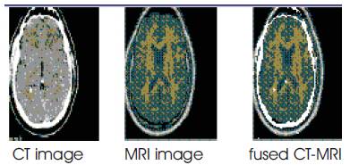
Figure-1
This method is also very useful to the display of teeth, since CT images cannot give good images of the relationship between teeth and the soft tissue around them. So we tried to fuse CT image and MRI image to solve this problem. Figure 2 shows the result of fusion.
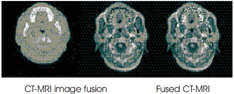
Figure 2
Subheading1
Conclusion
The routine is fast, semi-automatic and computationally inexpensive. In addition, the algorithm is extensible to 3-D. This is done using Java and makes the process quite easy in the hands of the medical staff. However, the technique is open to improvement in the near future so that we can compare and combine this with other registration-fusion algorithms, such as the Maximization of Mutual Information. In addition, the algorithm is extensible to 3-D. As a result, the clinician will have the opportunity to choose among different software registration-fusion techniques.