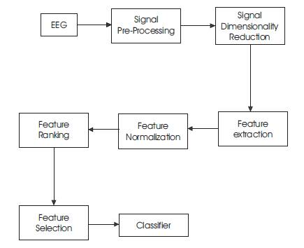
Figure 1. Block Diagram for the Proposed Work
In biomedical signal analysis, classification plays an important role and gives the promising solution to the electroencephalogram (EEG) analysis. An automatic EEG signal classification is proposed in this system and contribution of the diagnostician is replaced by using the soft computing techniques, since the manual classifications carried out in the clinical analysis is a time consuming task. In the proposed methodology, the EEG features are extracted from the raw EEG signals which are then fed to these ANFIS classifiers. It is a sophisticated framework for the classification of the different brain states in the human brain by representing their experts based knowledge as an Adaptive Neuro Fuzzy Inference System (ANFIS). This algorithm has a capability to detect the two types of brain states, including dementia and encephalopathy. Finally, the tentative outcome of the results is expressed in terms of classification accuracy and improves execution. The analyses are demonstrated with the ANFIS algorithm to improve and enhance the performances in the MATLAB.
Biomedical Signal Processing (BSP) has been rapidly developing and there is an increase in complex biological process in a wide variety of areas [8]. The biomedical signals help to determine and diagnose brain diseases and its types, but brain is a very complex organ in the human body [6]. The brain diseases are diagnosed by visual inspection of the brain signal and it is analyzed with the help of manifold learning model to explain about detailed information about the cerebral function of the brain. From the analysis of an anatomical structure of the brain, the treatment planning and the diagnosis of brain abnormality involves the standard manifold learning technique for the classification of different brain states. But, the existing physical classification of brain stages, especially brain diseases are time consuming and prone to error which leads to accuracy error [7]. The proposed Artificial Neural Network Fuzzy Inference System (ANFIS) for brain disease detection, which incorporates the best feature set and ANFIS rules to classify an abnormal brain signal for the corresponding brain state type results in better performance and improvements in the classifier system for classifying the normal and abnormal types of signal [10]. The ANFIS model is used for Brain signal classification to achieve better accuracy with large training sets and thus hybrid adaptive Neuro-fuzzy logic technique has been developed to yield the average accurate classification rate. ANFIS in brain abnormalities signal classification based on statistical analysis are provided by the diagnostician and then the results are compared with expert's results which provides best ANFIS classifier and its performance metrics [12]. Finally, the proposed system achieves the significant feature subset for best training as well as a test pattern for the classification of brain patterns in human brain. This classification approaches are used to replace the classifier system instead of a technician and achieves significant results.
The rest of this paper is structured as follows. Section 1, gives a brief overview of related works. Section 2 describes the proposed work. Section 3 explains the EEG signal processing and its extraction.
As all the researchers expressed their EEG signal analysis differently. It is not an easy task to judge and give the promising solution to predict the brain disease earlier. With the rapid development of advanced signal processing in the medical field, more and more researchers are yet trying to get the best solutions. The related works on EEG signal analysis proposed by various researchers are as follows:
Gurdeep Singh and Gautam Kaushal et al. [11] presented a comparative study of de-noising EEG signal using various methods such as Principal Component Analysis (PCA), Independent Component Analysis (ICA), and wavelet methods. On the basis of Signal – to - Noise – Ratio (SNR) and Mean Squared Error (MSE) values, wavelet transform has the better results to de-noise the EEG signal.
Abhinav Dixit and Swatilekha Majumdar [2] presented a comparative analysis between the Coiflets and daubechies wavelet filters on image based de-noising technique. Also they discuss the performance evaluation parameter like MSE, Peak Signal - to - Noise Ratio (PSNR) and SNR. From these evaluation parameters, they have concluded that Coiflets filter provided a better de-noising than the Daubechies filter [13].
Geeta Kaushik et al. [4] presented a detailed analysis of the de-noising and compression process of various wavelet families and biomedical signal for removing the noise from the signal and even compared their statistical parameter to find out the best result.
Tapan Gandhi et al. [3] have presented to find the most suitable wavelet family among all the existing families for the EEG signal analysis. These studies are used to classify the normal as well as abnormal elliptic EEG classifications. From this study, it was found that Coiflets 1 is the most suitable family among the wavelet families considered in this study for accurate classification of the EEG signals
G. Lalli et al. [6] proposed a new algorithm for feature extraction and selection. After that, the selected feature values are trained and tested for the different classifiers. From this study, ANFIS with subtractive clustering are better for the EEG signal classifications.
G. Lalli et al. [5] presented a selection of the best five feature values from the retinal images. After that, it underwent the ANFIS training, testing, and checking process for the calculation of the classifier accuracy.
The process of EEG signal analyses involves identifying the artifacts in the signal according to the specialist, reduction technique and classifier, which represents the EEG profile. The following block diagram (Figure 1) involved in the automatic brain stages classification to obtain the accurate result has been illustrated. The EEG data which have been collected from the hospitals are analyzed through several procedures, including signal processing, feature extraction, feature selection, and Neuro-fuzzy classifier. The primary objective of this work is to select a most suitable wavelet function which is used to remove the system or device artifact from the raw EEG signals. This is followed by the wavelet based signal decomposition. By this way, the secondary objective is achieved by performing the feature extraction and selections [9]. Finally, the selected features are fed to the classifier system to analyze the performance metrics.
Figure 1. Block Diagram for the Proposed Work
Raw EEG data are processed in two steps, namely conversion process and pre - process. Initially, the signal samples are stored in the form of text and then the stored texts are again converted into a signal. This process is called as a conversion process. In order to provide a valid input for programming in MATLAB, the signal conversion process is very much essential. Followed by the conversion process, pre-process is performed. For the pre-processing method, the de-noising technique is used.
Though, the research work has been done till date in the area of EEG signal de-noising, the problem has occurred till now. From the variety of approaches studied, the wavelet transform is found to be an effective timefrequency analysis tool for analyzing the EEG signals [1]. Several wavelet families are available for de-noising the EEG signals. Depending upon the types of artifacts, the wavelet is chosen according to the convenience and the requirement of the applications. This process is carried out by using the Daubechies family of order 4 for removing the system artifacts. On the basis of various evaluation parameters like MSE, PSNR, SNR calculated by different authors it is concluded that the wavelet method gives the best de-noising result with its multi-resolution capabilities. The wavelet transforms are used to analyze the signals in both time and frequency domain. It is also used to remove low noise amplitudes from the signals by the selection of the best wavelet family and its order. In this study, the Daubechies wavelet family of order four has been used to remove the noise present in the signals [4]. The de-noising has been classified into a three steps, namely signal decomposition, thresholding, and signal reconstructions. They are as follows:
A wavelet type and its order are chosen and decomposed up to level l.
For every level of decomposition, the selection of threshold value is an essential one and then it is followed by applying the thresholding in detailed coefficients.
The reconstruction is calculated based on the approximation coefficients of level l and the modified detail coefficients of levels from 1 to l.
The selection of the Daubechies wavelet of order four is on the basis of various evaluation parameters like MSE and PSNR.
The wavelet decomposition provides a time-frequency illustration of the EEG signals. The outputs of the de-noised signals are in time domain only, but there is a need of signal analysis in both the time and frequency domains. So, the decomposition is performed after the selection of wavelet family, order, and its decomposition level. This section explains the Haar wavelet decomposition level of 1. After applying the decomposition, the resultant signals are split into higher and lower frequency components from the merged one. This method is a basic step for the extraction method and provides the input for the classifier.
Signal dimensionality reduction is an important step in EEG data analysis. It can improve the learning efficiency and also improves the prediction performance. In this paper, signal dimensionality reduction is carried out with two well-known methods, namely feature extraction and selection. The feature extraction is used to reduce the number of random variables under some consideration. The extraction is one of the sub-processes of the reduction technique. On the basis of some statistical calculations, these reductions are achieved. The calculation involves the following steps, namely calculation of total number of sample values per head and the selection of dimensionality reduction variation values from minimum 4 to maximum 6. Finally, from the basis of the reduction values, the extraction and selection step is carried out. The aim of feature extraction is to identify the various patterns of brain activity and also to be used as input to a classifier. The performance of the recognition system depends on the feature extraction, selection, and the classification algorithm. In this method, the redundant features are removed from the decomposed signals by using the adaptive filter model. These extracted features are the fundamental basis for the detection, classification, or regression tasks in biomedical signal processing and is one of the key steps in the data analysis process. These features help in expressing a data into a new form. The extracting features from the decomposed signals are called as a probability co-variance. In this way, the redundant features are removed and the computational cost is improved by the adaptive filter model. Although there are more methods proposed for EEG extraction in the literature [3, 5]. This algorithm is chosen because, the adaptive filter has a capability to separate the large variation sample values from the input signals. It also, shows better results than other methods. The next step involves the feature normalization and feature selection based on their ranking. The linear discriminant analysis algorithm is developed to select the best features. These algorithms are the basic preprocessing step for the pattern-classification, machine learning applications, and also commonly used reduction technique. The algorithm is performed by the following steps:
Step 1: Formulation of the Eigen vectors from the extracted EEG data /sample set is done and called as scatter matrices.
(i.e., between the class matrix and within the class matrix).
Step 2: Perform the Min – Max scaling between the scatter matrices from the following equations is done.
Step 3: Sorting the Eigen vector from high to low (ascending order)
Step 4: The best five features from the sorting Eigen vector (1:5) is selected.
In the advent of the modern medical systems, there is a need for the model of expert knowledge based system according to input and output data given to it. The data could be obtained from the expert's knowledge or experimental analysis in the form of per fect mathematical modelling. Due to certain difficulties in manual analyses of these data, a suitable approach is essential in order to identify the relative information about the system. In such clinical situations, the earlier diagnosis and prognosis are the difficult task nowadays. So, the soft computing techniques have been used to address these problems and explore their knowledge as a set of IF-THEN rules. That can be helped to easily understood by people without the clinical expertise. The main focus of this research is to develop a novel approach based on the fuzzy inference system in modeling survival of Encephalon patients using recognized prognostic factors. While aiming to clearly classify the normal, abnormal EEG classifications and also expecting to obtain maximum accuracy of results. The adaptive Neuro-fuzzy inference system can be easily understood by clinicians and assists them to predicting the individual features and planning for further treatments. ANFIS is a combination of soft computing techniques, such as fuzzy inference system and neural network. This technique used to test the trained membership functions can be with better accuracy, rather than the clinical analysis. It is an efficient tool for handling the complicated system, which consist of linguistic information and data information simultaneously. The algorithm is based on the hybrid learning algorithm, which have a third type of inference system. From the basis of prognosis, a hybrid intelligent system is developed that is going to represent clinical expertise knowledge called as an Adaptive Neuro-fuzzy Inference System (ANFIS). Finally, the extracted knowledge can be represented into an IF and THEN rules. The above assumptions can be achieved by their extracted rules and have to be evaluated in terms of average time complexity computation time (in sec), average classification error and performance accuracy by using the ANFIS with subtractive clustering. This approach is going to attain a better balance between the training and testing stage.
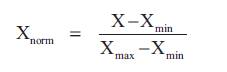
Adaptive Neuro-fuzzy Inference System is one of the soft computing approaches constructed by using a given set of input and output. The membership functions and rules of these systems are adjusted by using these algorithms to make them learn from the expert’s data and they are modeling. The classifier model is constructed by using two sets of fuzzy rules based on a Sugeno model. Fuzzy Logic and Neural network have been widely used as signal classification tools in biomedical applications and provide a high accuracy rate. The main difference between the normal and abnormal signals is the changes in frequency component that are provided by the experts which are used in classification system in the way of statistical analysis and are expressed in numerical quantitative method. The ANFIS systems implemented in the paper takes the selected features from the feature vector is obtained from the EEG signals as the input patterns and are classified into different brain stages.
One of the possible ways of modeling the ANFIS is the ANFIS architecture and it will be constructed by using the set of fuzzy rules that are shown in the following Figure 2.
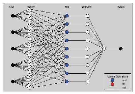
Figure 2. ANFIS Structure
The following statistical measures as in Figure 3 were used to analyze the performance of the classifier system. True Positive (TP) is the number of correctly classified abnormal signal, True Negative (TN) is the number of correctly classified normal signal, False Positive (FP) is the number of incorrectly classified abnormal signal, and False Negative (FN) is the number of correctly classified normal signal. Sensitivity is the proportion of the number of TP decisions to the number of actual positive cases (TP+FN). Specificity is the proportion of the number of TN decisions to the number of actual negative cases (TN+FP). Total correct classification (TCC) is the proportion of the number of correctly classified decisions (TN+TP) to the number of all cases (TN+FN+TP+FP).
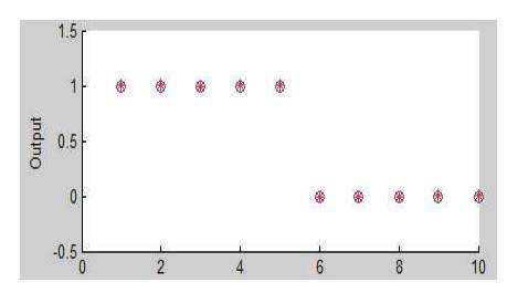
Figure 3. Performance Test between Training and Testing Membership Function
The paper demonstrates the implementation of automatic EEG signal classification based on Neuro-fuzzy classifier. The neurobiological disorders EEG signals are available on the medical database that are provided by the technician have been utilized for this study. For this study, EEG signals have been taken into consideration. The recording contains the following electrode positions F, N, O, S, and Z each sampled at 100 Hz. The EEG signals used from the database are first carried out in the conversion step followed that the signals are preprocessed by filtering the whole data. The data is filtered using de-noising technique. This filtering step removes the unwanted artifacts from the EEG data and improves the classification accuracy. In this work, wavelet based denoising is used. For the consideration of convenience, accuracy and computational time, few wavelet families are considered and are shown in the following Table 1. Table 1 depicts the reason for selecting the wavelet family in a de-noising purpose.
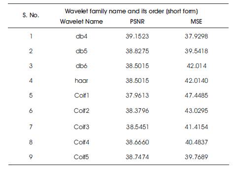
Table 1. Comparative Analysis Table for Different Wavelet Families
From this analysis, Daubechies family of order 4 provides a better fit to remove the system artifacts from the EEG signal. The de-noising technique used for this purpose gives the most promising technique for the noise removal from the EEG signals for diagnostic classification. The variations in the EEG patterns with respect to the brain stages are very subtle and therefore require advanced signal processing techniques to extract the features for brain state classification. The Weiner filter techniques are preferred for feature extraction as, owing to its best to separate the variation between these features and it can deal with the non stationary, complex and dynamic nature of the EEG signals. After that, the extracting values from the decomposed signals and the redundant features are removed from this process. Also selection of features values are performed by the LDA algorithms as mentioned above. To solve this problem, the removal of redundant feature has been employed to construct the features that correlate with EEG brain patterns and has the ability to reflect the difference between the brain stages. The features representing the brain stages are extracted by applying reduction technique on the decomposed signals and are used as inputs in Neuro-fuzzy based ANFIS classifier. The extraction technique is required to generate features for detection and discrimination for critical applications. The adaptive model is used to extract only the useful information from the signal about the process under study. For this work, the concept of adaptive filter technique has been used. It has the capability to automatically select the features that are highly correlated with the variations in the EEG signal with different brain stages. It works well with the small feature vectors. The relative features are extracted from the generating features and gives a best feature vector of 5 features. Moreover, there are no further selection values in the data set. Hence, the performances of the classifier are evaluated on the basis of accuracy. This helped in obtaining lesser computational time for the system. The normalized feature vectors representing EEG signals were used to create the training and test data set for the ANFIS classifier. Five sets of feature vectors with targeted outputs corresponding to 2 brain stages were constructed for training purposes. The training input set was formed by taking 10 patterns of 5 features each where each pattern represented per brain stage to the ANFIS classifier as training data with targeted output. The ANFIS classifier designed using the mentioned architecture details is then tested using a total of 10 patterns of 5 features each, which corresponds to the classification stage outputs obtained from the ANFIS classifier. The final outputs were then used to generate the variations in the brain stages obtained from the EEG experts. The justification for using this approach is that it reduces the computational time of the classification system and improves their performances.
These evaluations are used to find out the trade-off between the machine learning approach and the clinical analysis. Though the accuracy of the classification was high and computational time was low were found to be the best for EEG signal classifications, as shown in Table 2. It is clearly evident that the proposed approach provides a much higher accuracy as compared to clinical approaches. Owing to the suitably, high accuracy is achieved using the Neuro-fuzzy technique. The methodology presented in this paper can be utilized for designing automatic brain stage classifiers, which can be used by the neurologists for detecting various brain disorders.
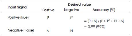
Table 2. Performance Measure of the Classifier
In this work, the appropriate wavelet families are selected for the de-noising and decomposition techniques. Sixteen features are extracted from the decomposed waveforms, after which, each extracting features are selected by the linear discriminant algorithms and are used to classify the cases. ANFIS with subtractive clustering is found to be the best classifier for analyzing the EEG signals with the more critical cases that resembles the accurate analysis of neurobiological disorder. Thus reducing the processing time and moreover the selection of the appropriate classifier through the performance analysis, leads to the improvement in the EEG signal analysis in comparison with the clinical analysis.
The authors are thankful to the authority of Kuppusamy Naidu Hospital, Coimbatore for giving access to their Encephalon disease related database and his support with experimental EEG signals used in their research.